
The brain is a very important part of your body and it should be properly taken care of. Regular checkups and scans will help you determine any disease or abnormalities in the brain, far before they become something serious. Brain scans can be used to diagnose various kinds of diseases, tumours, stroke, haemorrhage or even abnormal brain development. The most prominent scanning technologies are, CT, MRI, PET and SPECT.
Computed Tomography (CT scan)
CT scan uses X-rays to project a 2D image of the tissues, organs and bones. CT scans are very helpful for a proper diagnosis and can easily detect haemorrhage in people who have recently complained of brain strokes. Furthermore, early detection enables them to go for an immediate intravenous treatment to dissolve the clots.
CT scans also aid in understanding how the brain works and detect any anomaly in it. It usually takes about 20 minutes and is used for people who cannot go for MRI and X rays. However, CT scans are not suitable for pregnant women as they may pose a potential threat to the foetus.
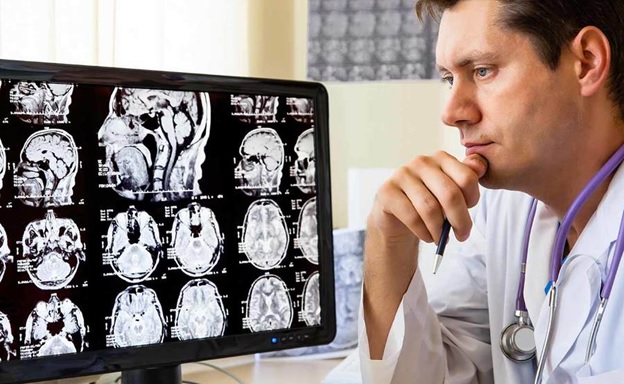
Magnetic Resonance Imaging (MRI scan)
MRI uses a powerful magnetic field and a computer-generated radio wave, which is able to produce a detailed image of the tissues. It uses different sequences of a magnetic pulse to show anatomical images of our brain and spinal cord. It can also measure the rate of blood flow and reveal in-depth figures about iron deposits and other minerals in our bodies. MRI can be used to diagnose stroke, spinal cord and brain tumours, a traumatic brain injury, infection, inflammation or all types of brain damage.
People who have a pacemaker or other implanted device should not go for MRI as the strong Magnetic fields can interfere with its proceedings and cause serious issues. Furthermore, we only recommend consulting specialists such as Inside Radiology to be assured that the tests are conducted in a proper and secure way.
Positron emission tomography (PET)
PET scans can give a 3D or 2D image of your brain’s activity levels by tracking the movements of a radioactive isotope, which is injected into the bloodstream before the PET. It facilitates the detection of brain tumours, slow blood flow or diseased tissue. PET scan is a follow-up scan after CT or MRI, which helps in evaluating the specific affected parts of your brain. It is a short process where a low-level radioactive isotope known as the tracer is injected into a person’s bloodstream. By using different compounds, more than one type of brain function can be traced simultaneously.
Single Photon Emission Computed tomography (SPECT)
SPECT is a nuclear imaging test, which can be used to evaluate certain brain functions. Alike PET, a radioactive isotope is injected into the body. It can also be a follow-up scan to an MRI to diagnose infections, tumours, degenerative diseases, and seizures. This can also be used to diagnose Parkinson’s disease with the help of dopamine transporter. The gamma camera rotates and provides a detailed 3D image of the freely-moving tracers in the brain.
Electroencephalography (EEG)
EEG is a way of monitoring a patient’s neuron activities through the skull. It is used to diagnose infectious or inflammatory disorders, which may affect the human brain. Sleep disorders’s diagnoses or a precision-requiring test of brain activity levels can be done through EEG.
It is a painless and risk-free test, where the person is made to sit in a chair and some cups of electrodes are placed on their head with a conducting paste. The signals are converted into appropriate data by the machine. During an EEG session, an external stimulus is given to see how the brain reacts to it.
We hope his article has helped you understand the various neurological tests that can be conducted to detect brain problems.